md-medicaldata
Main menu:
- Naslovna/Home
- Arhiva/Archive
- Godina 2023, Broj 3
- Godina 2023, Broj 1-2
- Godina 2022, Broj 3
- Godina 2022, Broj 1-2
- Godina 2021, Broj 3-4
- Godina 2021, Broj 2
- Godina 2021, Broj 1
- Godina 2020, Broj 4
- Godina 2020, Broj 3
- Godina 2020, Broj 2
- Godina 2020, Broj 1
- Godina 2019, Broj 3
- Godina 2019, Broj 2
- Godina 2019, Broj 1
- Godina 2018, Broj 4
- Godina 2018, Broj 3
- Godina 2018, Broj 2
- Godina 2018, Broj 1
- Godina 2017, Broj 4
- Godina 2017, Broj 3
- Godina 2017, Broj 2
- Godina 2017, Broj 1
- Godina 2016, Broj 4
- Godina 2016, Broj 3
- Godina 2016, Broj 2
- Godina 2016, Broj 1
- Godina 2015, Broj 4
- Godina 2015, Broj 3
- Godina 2015, Broj 2
- Godina 2015, Broj 1
- Godina 2014, Broj 4
- Godina 2014, Broj 3
- Godina 2014, Broj 2
- Godina 2014, Broj 1
- Godina 2013, Broj 4
- Godina 2013, Broj 3
- Godina 2013, Broj 2
- Godina 2013, Broj 1
- Godina 2012, Broj 4
- Godina 2012, Broj 3
- Godina 2012, Broj 2
- Godina 2012, Broj 1
- Godina 2011, Broj 4
- Godina 2011, Broj 3
- Godina 2011, Broj 2
- Godina 2011, Broj 1
- Godina 2010, Broj 4
- Godina 2010, Broj 3
- Godina 2010, Broj 2
- Godina 2010, Broj 1
- Godina 2009, Broj 4
- Godina 2009, Broj 3
- Godina 2009, Broj 2
- Godina 2009, Broj 1
- Supplement
- Galerija/Gallery
- Dešavanja/Events
- Uputstva/Instructions
- Redakcija/Redaction
- Izdavač/Publisher
- Pretplata /Subscriptions
- Saradnja/Cooperation
- Vesti/News
- Kontakt/Contact
 Medicinski fakultet
 Pasterovo društvo
- Disclosure of Potential Conflicts of Interest
- WorldMedical Association Declaration of Helsinki Ethical Principles for Medical Research Involving Human Subjects
- Committee on publication Ethics
CIP - Каталогизација у публикацији
Народна библиотека Србије, Београд
61
MD : Medical Data : medicinska revija = medical review / glavni i odgovorni urednik Dušan Lalošević. - Vol. 1, no. 1 (2009)- . - Zemun : Udruženje za kulturu povezivanja Most Art Jugoslavija ; Novi Sad : Pasterovo društvo, 2009- (Beograd : Scripta Internacional). - 30 cm
Dostupno i na: http://www.md-medicaldata.com. - Tri puta godišnje.
ISSN 1821-1585 = MD. Medical Data
COBISS.SR-ID 158558988
АКТУЕЛНОСТИ У ХИСТОЛОГИЈИ И ЕМБРИОЛОГИЈИ
„NEWS IN HISTOLOGY AND EMBRYOLOGY”
Author
dr Dejan Miljković
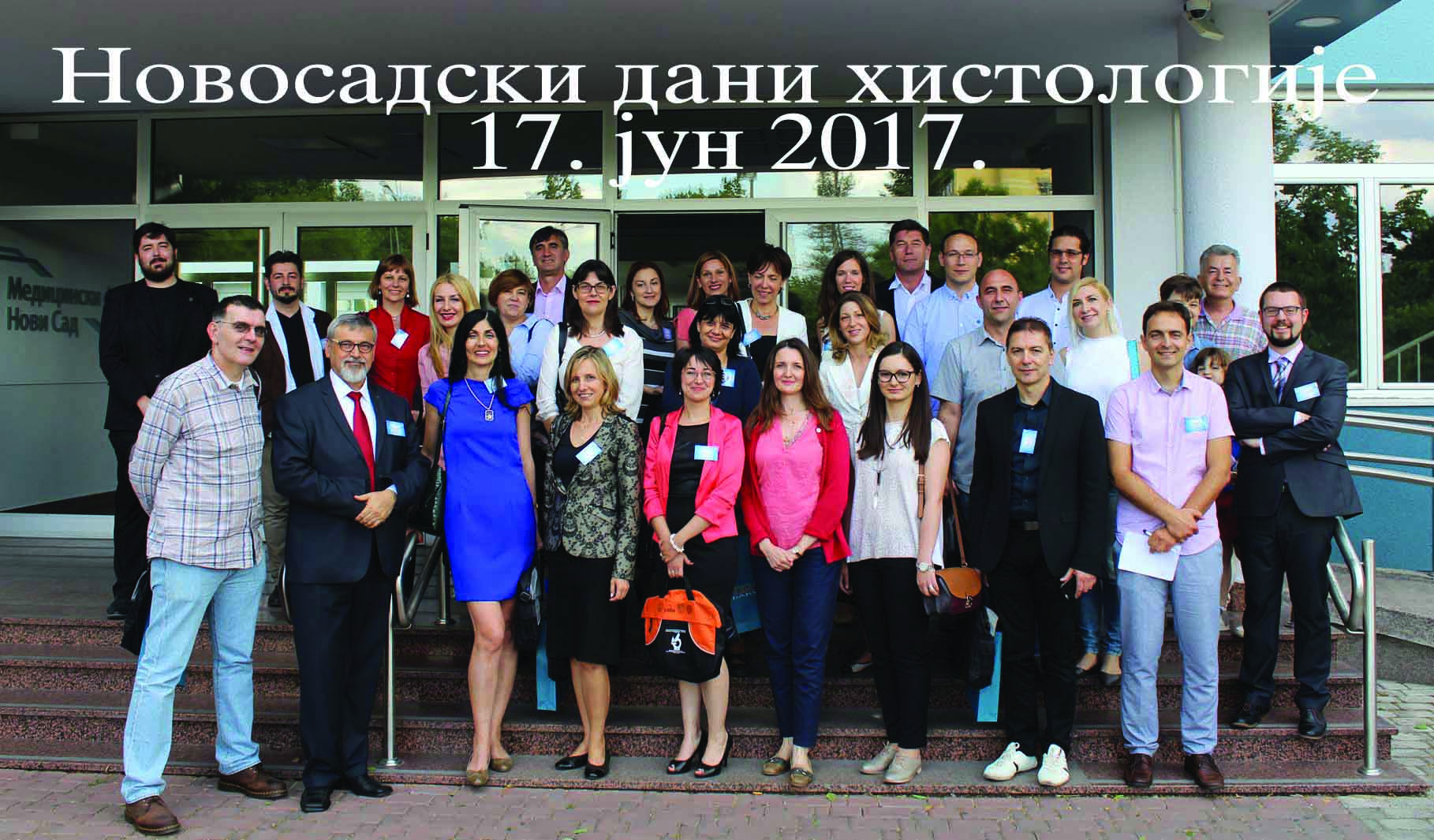
U subotu, 17. juna 2017. godine sa početkom u 10 časova, posle više od 25 godina Medicinski fakultet Univerziteta u Novom Sadu obeležio je reosnivanje Zavoda za histologiju i embriologiju. Svečanosti su prisustvovali, pored domaćina − dekana, prof. dr Snežane Brkić, prodekana za nastavu prof. dr Zorana Komazeca, prodekana za akreditaciju i kontrolu kvaliteta, prof. dr Biljane Srdić Galić, prodekana za specijalizacije, prof. dr Milice Medić Stojanoske, prodekana za nauku prof. dr Duška Kozića, direktora Centra za medicinska i farmaceutska istraživanja i kontrolu kvaliteta, prof. dr Biljane Božin, šefa Zavoda za histologiju i embriologiju, doc. dr Ivana Čapo, šefa Katedre za histologiju i embriologiju, prof. dr Dušana Laloševića, i gotovo svih članova Katedre za histologiju i embriologiju i Katedre za patologiju, kao i kolega sa velikog broja drugih katedri i stranih gostiju, prof. dr Marius Raica, rektor i šef Katedre za histologiju i embriologiju Medicinskog Univerziteta u Temišvaru i prof. dr Anca Maria Cimpean, kao i predstavnici Katedri za histologiju i embriologiju Medicinskih fakulteta iz Beograda, Kragujevca, Niša, Prištine-Kosovske Mitrovice, Vojnomedicinske akademije u Beogradu, Stomatološkog fakulteta u Beogradu i Veterinarskog fakulteta u Beogradu.
Obeležavanje ovog značajnog jubileja nastavljeno je stručnim sastankom – simpozijumom pod nazivom „Aktuelnosti u histologiji i embriologiji – Novosadski dani histologije i embriologije”. U okviru skupa održana su predavanja na aktuelne teme u histologiji i embriologiji, a predavači su bili predstavnici katedri za histologiju i embriologiju iz cele Srbije, kao i gosti sa Katedre za histologiju i embriologiju Medicinskog Univerziteta u Temišvaru.
Author Prof. dr Dušan Lalošević
Tokom prvih 30 godina, Medicinski fakultet u Novom Sadu, osnovan 1960, imao je čuveni Zavod za histologiju i embriologiju koji je osnovao akademik Radivoj Milin. Poznat kao odličan predavač i organizator, prof. Radivoj Milin je izabrao odlične saradnike koji su radili histohemiju i dijagnostičku elektronsku mikroskopiju. Međutim, 1991. Katedra za histologiju i embriologiju ostala je bez svoje osnovne nastavne baze, Zavoda za histologiju i embriologiju Medicinskog fakulteta takozvanom „integracijom“ sa Centrom za patologiju Kliničkog centra Vojvodine, a zapravo preuzimanjem zaposlenih sa Zavoda za histologiju i embriologiju. Iz želje da se rad poboljša, na žalost, došlo je do zaustavljanja naučnog rada iz histologije, jako otežanog nastavnog rada, jer je nastavnika i saradnika na Katedri bivalo sve manje, a pri tome su nastavnici i saradnici histologije i embriologije bili bukvalno preopterećeni rutinskim radom na patologiji KC Vojvodine. U jednom periodu Katedra je imala samo jednog nastavnika, zatim dva u trećinskom radnom odnosu, što je daleko manje nego ranije i nego što imaju druge slične katedre našeg fakulteta. Zbog poznate situacije u zdravstvu, Centar za patologiju i histologiju KCV, ima histologiju samo u nazivu, ali nije u mogućnosti da radi kao nastavna baza Katedri za histologiju. Na patologiji svaki uzorak se posebno fakturiše Fondu za zdravstveno osiguranje i tu jednostavno nema prostora, a ni osoblja za neki naučni rad iz histologije. Nakon 25 godina zahvaljujući razumevanju uprave fakulteta, uspeli smo da ponovo osnujemo Zavod za histologiju i embriologiju, za sada sa četiri zaposlena istraživača. Kvalitet njihovog eksperimentalnog rada i novih publikovanih radova obećava kvalitetan razvoj novog Zavoda
Founded in 1960, Faculty of Medicine in Novi Sad had famous Institute of histology and embryology. Founder of Institute, academician Radivoj Milin, elected excellent collaborators who carried out histochemistry and diagnostic electron microscopy. But, at the beginning of year 1991, Department of histology and embryology at the Faculty lost its own fundamental basis, Institute of histology and embryology. Because of so called „integration” with Institute of Pathology, Clinical Centre of Vojvodina, histology as scientific discipline lost possibility for experimental work and development. Especially hard situation has become with teaching staff of histology and embryology, who worked as clinical pathologists, on enormous number of autopsy and surgical bioptic material. Twenty-five-years after, thanks to understanding of management of Faculty, we refounded Institute of histology and embryology, now with four scientific staff that work only at the Faculty. High quality of current experimental work and newly published papers indicate that future development of Institute will be sure.
Author Prof. Dr. Marius Raica
In last 150 years, histology was a basic discipline of medical education. Since the invention of the microscope in the XVII century until now, histology was thought to be the microscopic anatomy. On one hand this is true, as it deals with normal structure of cells and tissues, but on the other hand it is more connected to the pathology and particularly involved in the formulation of diagnosis. Nowadays there an obvious tendency to have a histology that is strongly connected to function of tissue and organs and moreover, to clinical application and involvement of the normal tissues in pathological processes. Teaching of histology evolved along the years from a one-way transmission of data – namely descriptive histology, to interactive lectures, IT supported practical works, and computer-assisted students’ evaluation. Research in histology is mainly connected to pathological conditions, despite at present time a full molecular profile of normal tissue is not available. What the future will bring for Histology? It is not easy to make predictions, particularly about the future! It is expected that soon the microscope will be replaced by computer-assisted microscopes in the labs for student. In such a way teaching and learning could be done at any time and from anywhere. 3D and 4D of cells and tissues are very close and their routine application in Histology will be ready no later than 2025. In the near future, we can suspect a very important role of histologists in research, as pathologists will be more and more involved to refine the diagnosis. Besides all these, most probably the core and basic Histology is appropriate for the training of future scientists. Author Prof. Dr. Anca Maria Cimpean, MD, PhD
The beginning of histology overlapped with the start of medical university in Timisoara. Moreover, the first rector of the university, professor Serban Bratianu, was also the first head of the histology department. Today, after 77 years, this history repeats itself: the medical school in Timisoara is led by a histologist too! The work with students and research came together in our department, professor Bratianu having several contacts with famous scientists from outside, especially with those of french medical school. More than 10 articles of professor Bratianu are mentioned on PubMed, unfortunately not those from period spent in Timisoara. Some ideas of professor Bratianu as immunity in cancer or viral involvement in carcinogenesis are considered nowadays as revolutionary topics in the field. Life forced professor Bratianu to move to Lassy and histology was really “governed” by women who were mainly focused on teaching except professor Maria Dragan who, somehow,revigorated research which continues today. Slowly, present histology in Timisoara turns to become more than a preclinical subject being focused on multidsciplinary approach of normal and tumor tissues from microscopic to molecular level. Angiogenesis Research Center and Lab of Molecular Pathology are two of the newest part of histology in Timisoara organized by the present head of the histology department.
Beyond appearances, each member of the histology department hides some funny life stories, most of them related to their way through histology or their extracurricular activities. Together with the serious part of it, histology from Timisoara will be described here as being scenic and full color histology beyond the lens of the microscope.
Author Prof. Dr. Anca Maria Cimpean, MD, PhD
Baby Hamster Kidney Fibroblasts (BHK 21/C13) have the ability to preserve their properties when they are transfected with different viruses and thus, they are used mainly in experimental virology and vaccine production. Few data are available regarding the use of this cell line in experimental cancer research, excepting the observation of their high invasiveness. Their phenotype is not yet characterized and this may be the reason for their limited use as an experimental alternative for testing different anticancer therapeutic agents. We aim to characterize this cell line regarding growth factors expression and markers known as therapeutic targets in other malignancies and also to test the behaviour of BHK 21/C13 cell line derived tumors implanted on chick embryo chorioallantoic membrane (CAM) and treated with anti-podoplanin antibodies. Immunohistochemical assessement showed positive reaction for vimentin, CD117, smooth muscle actin (SMA), vascular endothelial growth factor (VEGF A) epidermal growth factor receptor (EGFR) and PROX 1 and negative reaction for platelet derived growth factor B (PDGF B), Neuron Specific Enolase (NSE), S100 protein, CD34, Ewing Sarcoma and podoplanin in tumor cells. Despite of its rapid growth,low to medium proliferative rate has been detected together with a low intratumor blood vessels density. Tumors growing from BHK 21/C13 cells implanted on CAM were sensitive to disodium cromolyn and anti-podoplanin antibodies, two therapeutic agents which produced a massive necrosis of implanted tumors. Despite controversial behaviour (high invasiveness versus medium proliferation rate and low microvessel density) observed in the present study, the immunohistochemical characterisation mentioned above represents the first step in revealing BHK 21/C13 cell line use as a reliable tool for experimental cancer research.
Author Doc. dr Aleksandar Mirčić
Microtubules play a role in a large number of cellular functions, including a crucial role in chromosome separation during the cell division. In the treatment of malignant tumors microtubule drugs are used, which stabilize or disassemble microtubules and perturb microtubule function.
We were investigated paclitaxel effects (which stabilize microtubules) on C6 glioma cell lines incubated 24-72 hours with drug. Cells were washed, fixed and processed for embedding in Epon. On semithin sections, we counted cells and the percentage of apoptotic, mulitinuclear and cells blocked in mitosis was determined and expressed as indexes, while ultrathin sections were contrasted and analyzed by TEM. We used primary monoclonal antibodies on α-tubulin and fluorescence secondary antibody (Alexa 488) for analysis by confocal microscope. By Western blot we analyzed the proteins involved in process of apoptosis and autophagy.
Paclitaxel induced significant increase of mitotic index (35%) after 24 hours incubation and especially multinuclear index (64%) after 72 hours, while apoptotic index was slightly increased in the first 24 hours incubation, after was decreased what was showed by TEM analysis. Microphotographs displayed bundles of microtubules, also multinuclear cells and after 72 hours incubation we noticed number of autophagosomes, and occasionally there were cells showing both apoptosis and autophagy. Confocal microscopy analysis clearly showed bundles of microtubules and plenty of nuclei in cells. Western blot analysis presented that proteins involved in apoptotic process were not expressed, while highly expressed LC3II protein displayed autophagy in cells.
Our results showed that incubation of C6 glioma cell lines with paclitaxel, by disrupting dynamics of microtubules, triggered different types of programmed cell death. In small amount of cells apoptotic features were observed, after 72 hours of incubation autophagy was dominant, what WB analysis confirmed. Great number of cells, blocked in mitosis, independent of apoptotic process escaped mitotic blockade and aberrantly finished mitosis, manifested with multinuclear cells.
Author Doc. dr Mila Ćetković-Milisavljević
Trigeminalna neuralgija se karakteriše iznenadnim, obično jednostranim, probadajućim, kratkim, intenzivnim bolovima, često kao odgovor na senzornu stimulaciju delova lica, u području inervacije grana trigeminalnog nerva. Cilj našeg istraživanja bio je da ispitamo strukturu trigeminalnog nerva i trigeminalnog gangliona, njihovu vaskularizaciju i vaskularne odnose koji mogu da imaju ulogu u razvoju trigeminalne neuralgije.
Istraživanje je obavljeno na 20 parova trigeminalih nerava (TN) i gangliona (TG) (ukupno 40 preparata, osoba oba pola 14 muških i 6 ženskih), bez znakova promena na strukturama centralnog nervnog sistema, starosti od 26 do 79 godina (prosečno 55,65). Materijal za histohemijske i imunohistohemijske metode bojenja je pripreman na standardan način.
Dužina cisternalnog segmenta TN kretala se od 10,2 go 20,4 mm (prosečno, 14,01 mm). Zona centralnog mijelina prosečno je iznosila 4,91 mm kod senzornog korena (od 3,1 to 10 mm) i prosečno 0,77 mm kod motornog korena (od 0,5 do 1 mm). Najčešći kontakt korenog dela trigeminalnog nerva bio je sa gornjom cerebelarnom arterijom (20%), sa petroznom venom (24%) i prednjom donjom cerebelarnom arterijom (12% slučajeva). Ektopični neuroni su pronađeni u 26 TN (18 osoba), odnosno u 65% nerava ili 90% osoba. Kod 8 (40%) osoba (prosečne starosti 52,2 godine) izmešteni neuroni su uočeni kako na desnom tako i na levom TN, u 10 (50%) osoba (prosečne starosti 58,2 godine) bilo u desnom ili u levom TN, a kod 2 (10%) osobe (prosečne starosti 56,5 godina) nije bilo ektopičnih neurona u TN. Identifikovali smo 73 ektopične nervne ćelije, 21 malu, 48 srednje veličine i 4 velike. Prosečan broj mastocita po kvadratnom milimetru preparata TG iznosio je 1,3, a kretao se od 0 do 6 ćelija. Prosečan broj mastocita po kvadratnom milimetru preparata dure sa srednjim moždaničnim sudovima iznosio je 6,8, a kretao se od 5 do 8 ćelija.
Rezultati našeg istraživanja podržavaju hipotezu o postojanju neurovaskulrnog kontakta između okolnih krvnih sudova i korenog dela TN i mogućeg uticaja na razvoj trigeminalne kompresije. Broj mastocita je daleko veći u periganglijskom duralnom prostoru u odnosu na tkivo TG.
Author Prof. dr Danica Marković
Upotreba stranih (veštačkih) materijala za obnovu tkiva u organizmu je poznata iz zapisa starih civilizacija. Opisano je korišćenje staklenih očiju, komadića drveta i kamena u sanaciji povreda kod ljudi i životinja. Zlato se u zubarstvu upotrebljavalo pre 2000 godina. U vreme prvog svetskog rata, u ratnoj hirurgiji su korišćeni materijali od padobrana u hitnim operacijama kod povreda mekih tkiva.
Napredak u proizvodnji poboljšanih materijala sastoji se u tome da se savremenom tehnologijom dobiju biomaterijali što sličniji prirodnom tkivu u koji se unose, te da tako proces reparacije bude podstaknut i što kraći, a da prihvatanje od strane organizma prođe bez štetnih efekata.
Danas je upotreba biomaterijala u sanaciji rana, opekotina i nekroza veoma prisutna u reparativnim i regenerativnim medicinskim procedurama. Novi biomaterijali za meka tkiva, pre nego uđu u kliničku primenu, po međunarodnim standardima za upotrebu u veterini, medicini i stomatologiji, moraju da prođu preciznu proceduru provere koja se u prvom redu odnosi na njihovu biokompatibilnost. U tom smislu standardom su predviđeni životinjski modeli koji se koriste u predkliničkim skrining metodama za in vivo upotrebu i procenu neškodnjivosti biomaterijala. Histološka evaluacija biološkog odgovora tkiva na implantate je polazna metoda u procenama pogodnosti za korišćenje određenog biomaterijala u živom organizmu.
Biomaterijal (hidrogel sa nanočesticama srebra) je napravljen na Tehnološko metalurškom fakultetu Univerziteta u Beogradu (Katedra za fizičku hemiju i elektrohemiju). Biomaterijali su implantirani u subkutis (koža leđa, paravertebralno), 16 pacova (Wistar soja, starosti oko 3 meseca), sa odobrenjem Etičke komisije Fakulteta veterinarske medicine. 7, 15 i 30 dana nakon implantacije materijal je sproveden i urađena su bojenja Hematoksilin eozin (H/E) i Azan (Az). Kvalitativna i kvantitativna analiza urađena je morfološkim i semikvantitativnim metodama gde se posmatrao biološki odgovor svih parametara tkiva (epitel, vezivno tkivo, masno tkivo, mišićno tkivo, krvni sudovi) oko biomaterijala i upoređivan je sa netretiranim, kontrolnim, uzorcima kože.
Author Prof. dr Goran Radenković
Neural crest cells (NCC) can migrate into different parts of the body and express their strong inductive potential. In addition, they are miltipotent and are able to differentiate into various cell types with diverse functions. In the primitive gut, NCC induce differentiation of muscular structures and interstitial cells of Cajal, and they themselves differentiate into the elements of the enteric nervous system (ENS), neurons, and glial cells, all required for normal peristalsis, a prerequisite for normal bowel function. It has been undubitably shown that disorders or losses of interstitial cells of Cajal (ICC) underlie many motility disorders. ICC develop by way of mesenchymal cell differentiation in the outer parts of the primitive gut wall around the myenteric plexus (MP) ganglia, with the exception of colon, where they appear simultaneously at the submucosal border of the circular muscular layer around the submucosal plexus (SMP) ganglion. However, at the end of embryonal period of development, c-kit positive cells are the first to appear around the inception of the MP ganglion, which are markedly more abundant and morphologically different from the mature ICC forms. Our assumption is that NCC induce mesenchymal cell differentiation into c-kit positive precursors capable of differentiating into ICC and smooth nuscle cells (SMC). C-kit positive precursors could represent a key impact factor regarding the final differentiation of NCC into neurons and glial cells and formation of myenteric ganglia, with neurons subsequently excreting stem cell factor (SCF) and other signaling molecules. Under the impact of SCF, a portion of c-kit positive precursors lying immediately around the ganglia differentiate into ICC, while the rest differentiate into SMC.
Authors Doc. dr Miloš Bajčetić, asist. dr Ivan Zaletel, asist. dr Jelena Rakočević
Since 2004, when the Reticulum (official portal for online learning) was established, and when the first blended course on histology and embryology was developed at the Belgrade University School of Medicine, more than 5,000 students have attended more than 30 different blended courses on different subjects. In order to improve the quality of education with the help of new educational technology biggest challenges were selection of appropriate Learning Management Systems (LMS) and improving pedagogical and ICT competencies of teachers.
Moodle LMS allows personalized and collaborative learning. It has different forms of synchronous and asynchronous activities very useful for medical education (Lesson, Quiz, Discussion Board, Big Blue Button, Feedback, Glossary, Chat, Wiki, Games, etc.). Moodle provides real time feedback and opportunity for peer assisted learning. It also has very powerful learning analytic tools. From the point of view of end users (medical teachers and students) Moodle is very intuitive and user friendly. Another very important thing was fact that Moodle is open-source web application.
In order to improve pedagogical and ICT competencies of medical teachers at Belgrade University School of Medicine we developed five-week blended course „Introduction to e-Learning and Learning Management Systems” as well as five-day workshop „Active learning in Medical education”. Main objectives were to raise awareness in integrating the IC Technologies within the traditional existing curriculum, to introduce the basic pedagogical principles of active learning and to enhance the use of LMS in the teaching process.
Authors Doc. dr Sanja Milutinović-Smiljanić1, prof. dr Vesna Danilović1, prof. dr Božidar Brković2
Bone histomorphometry allows the quantitative study of bone microscopic organization and structure, so it is gold standard for examination of bone regeneration.
Histomorphometric analysis of new bone formation after the use β-TCP/Col (beta-tricalcium phosphate with type I collagen) and hemocollagen in the alveolar ridge preservation.
Bone specimens were obtained from 40 healthy patients divided into two groups, 2 and 4 months of healing. All treated postextracted sockets were preserved with β -TCP/Col and hemocollagen (Septodont, France) after tooth extraction in the alveolar ridge of the esthetic zone. Bone specimens were subjected to Goldner΄s trichrome staining. Image acquisition and morphometric assessment were performed using a stereological software package (Leica Application Suite Leica Microsystems software). The following structural parameters were analyzed: bone area (mineralized and nonmineralized), connective tissue, bone marrow, bone graft substitute and blood vessels.
All the sections after the healing of 2 and 4 months contained newly formed mineralized bone. In all specimens, graft material particles surrounded by the connective tissue with blood vessels, fibroblasts and collagen fibers intermixed with newly formed bone tissue could be recognized. The total bone area was significantly increased after 4 months healing (50.97 ± 2.58%) compared to control (26.79 ± 5.72%). The areas of mineralized new bone formation, with trabecular connectivity, were significantly increased after 2 months of recovery (26.10 ± 1.98%) compared to control (4.07 ± 1.60%), and after 4 months of recovery (25.28 ± 1.98%) compared to control (11.89 ± 3.19%).
This study shows that the use of β-TCP/Col I and hemocollagen is effective material in preservation of alveolar ridge. It is associated with significant enhance in bone formation and mineralization, with no signs of inflammation after observation period of 4 months. Further research is needed.
Author Prof. dr Anita Radovanović
U okviru naših ispitivanja promena koje se javljaju na zglobovima pasa u toku starenja, ipitivali smo karakteristike zglobne hrskavice tibije i femura, kao i sinovijalne membrane radnih (policijskih) pasa rase nemački ovčar, starijih od deset godina, kao i mladih (uzrast 3-5 godina) pasa iste rase koji su dovedeni na eutanaziju na Fakultet Veterinarske medicine. Kriterijumi prema kojima su psi bili uključeni u ispitivanje bili su sledeći: istorija bolesti bez znakova hromosti, negativan radiološki nalaz kolenog zgloba, zgloba kuka i lumbosakralnog dela kičme na prisustvo osteoartritičnih promena, negativan nalaz sinovijalne tečnosti na prisustvo markera inflamacije procenjen na osnovu fizičko-hemijskog ispitivanja i prisustva serum amiloida A. Histološkom analizom uočeno je da sa starenjem dolazi do smanjenja debljine hrskavice tibije, smanjene zastupljenosti proteoglikana, hipertrofije hondrocita površinske i srednje zone hrskavice femura i ekspresije kolagena tipa X u nekalcifikovanoj hrskavici. To je ukazalo na postojanje degenerativnih promena koje nisu narušile funkciju hrskavice, ali predstavljaju preduslov za moguću pojavu osteoartritisa.Takođe, dugotrajno fizičko opterećenje, kome su radni psi bili izloženi u toku svog radnog veka, nije dovelo do značajnih strukturnih promena u sinovijalnoj membrani. Povećanje broja kratkih i prstolikih nabora odgovara promenama koje su povezane sa starenjem, dok se povećanje broja slojeva ćelija u intimi može dovoditi u vezu sa adaptivnim odgovorom sinovijalne membrane na fizičko opterećenje. Na osnovu dobijenih rezultata može se zaključiti da umerena fizička aktivnost ima pozitivan uticaj na očuvanje strukture hijaline hrskavice i sinovijalne membrane kolenog zgloba tokom starenja.
Author Doc. dr Tamara Kravić-Stevović
Transmission electron microscopy (TEM) has been receiving extensive attentionin nanomedicine. Nanoparticles routinely used for nanomedical applications are carbon nanoallotropes: fullerenes, carbon nanotubes, and graphene. Nanoparticles are particulate materials with at least one dimension in the range of 1−100nm. TEM reveals both the presence of nanoparticles in cells, and favorable and unfavorable effects of nanoparticles on the ultrastructural features of cells treated with them. With the use of TEM graphene quantum dots (GQD) were visualized as graphene sheets with lateral dimensions less than 100nm. We have demonstrated the immunomodulatory and cytoprotective effects of large GQD in a mouse model of immune-mediated liver damage.Nano-scale size and a possibility for diverse surface modifications make GQD an indispensable nanostructured material. We have recently reported anticancer activity of photoexcitated curcumin nanoparticles and demonstrated that the irradiation with blue light significantly enhances the cellular uptake of nanocurcumin, leading to activation of c-JunN-terminal kinase, mitochondrial depolarization, and induction of apoptotic death in cancer cells. TEM images showed that the average size of nanocurcumin was 200nm, and that irradiated nanocurcumin penetrated the cell membrane and induced mitochondrial disintegration in cells in vitro. TEM has a significant role in nanomedicine in evaluation of both the presence of nanoparticles in cells and possible ultrastructural damage of cells.
Author Doc. dr Nela Puškaš
The hippocampal formation has the key role in the formation of memory and processing of emotional information. Also, it is a brain region involved in the regulation of hypothalamic-pituitary-adrenal axis and stress response. While the acute stress induces rapid adaptive responses, chronic exposure to stress leads to the overproduction of stress hormones and overactivity of HPA axis, which further leads to morphological and functional alterations in the brain, and finally to development of mood disorders, primarily depression.
Therefore, over the past few decades different animal models of depression were developed, based on the exposure to different types of repeated acute and/or chronic stressors, or intraperitoneal application of corticosteroids. Recently, it was shown that repeated administration of anabolic androgenic steroids results in changes indicative for a depressive state in normal rats too. On the other hand, it is well known that exercise has beneficial, anxiolytic effect on behavior in humans and animals.
Consequently, our focus was mainly on GABA-ergic interneurons in the hippocampus. Preliminary results of our experimental protocols, together with other previous studies, have revealed that the hippocampal GABAergic interneurons, mostly parvalbumin-positive (PV+) and neuropeptide Y-positive (NPY+), represent an especially vulnerable population of neurons in chronic stress. Immunohistochemical expression of NPY and PV generally decreases in the hippocampus in animals exposure to different stressors compared to control, but intensity varies depending on the experimental procedure and/or the exposure time. Similarly, exercise which has anxiolytic effect increases number of PV+ or NPY+ interneurons in the hippocampus. Also, changes were noticed in the DG, in the number of doublecortin-positive (DCX+) cells, which represent new born neurons. Namely, in different experimental protocols the number of DCX+ cells could decrease, indicating influence of stressors to adult neurogenesis, and vice versa, exercise could increase the number of DCX+ cells.
Author Asist. dr Dejan Miljković
Aristolochic acids are considered to be one of the main risk factors responsible for the occurrence of diseases such as aristolochic acid nephropathy (AAN) and possibly Balkan endemic nephropathy (BEN), especially aristolochic acid I (AAI). After many years of research, in experimental animals has been found that aristolochic acids are extremely nephrotoxic and carcinogenic compounds.
Our experiment included 64 NMRI mice that were divided into three groups: experimental group (n=32), vehicle control group (n=16) and control group (n=16). All animals were treated i.p. daily for seven days. Experimental group received AAI (10 mg/kg body weight). Vehicle control group recieved 2,5% saline solution of PEG 400 and control group received saline only. Before sacrifice, a 24-hours-urine collection was performed on days 8, 17, 29 and 59 in metabolic cages. Eight mice per experimental group and four mice per vehicle control and control group were euthanized on days 9, 18, 30 and 60. All mice were dissected individually and intracardiac puncture of the right ventricle was used to collect the blood. Biochemical parameters of urea, creatinine, uric acid, total protein and albumin in the blood and urine were used to confirm the damage of renal tissue.
By the morphometric and immunohistochemical analysis of kidney cortex of every experimental animal, it is expected that tubulointerstitial injury (acute tubular necrosis and mononuclear cell infiltration) differs qualitatively and quantitatively after 9 days, 18 days, 30 days and 60 days from the beginning of the experiment. A number of structural elements of renal glomeruli (capillaries, podocytes and mesangium) and tubulointerstitial injury in experimental animals should be different compared to control animals. Also, there should be a high correlation between morphometric histopathological data and biochemical parameters.
Author Doc. dr Ivan Čapo
Structural integrity of the basement membrane is necessary for proper migration of the neurons. Collagen is one of the key structural units of the basement membrane, and its synthesis is dependent on vitamin C. Based on the fact that neither humans nor guinea pigs are able to synthesize vitamin C we created a unique animal model in which prenatal vitamin deprivation in guinea pig fetuses led to a collagen synthesis disturbances, weakness, and finally a breach of pial basement membrane. The experiment included control (21) and vitamin C deprived- experimental group (21) of guinea pig fetuses. In experimental group pregnant guinea pig were deprived for vitamin C from 10th to 50th day of gestation. In each group we euthanized their pups at 50th fetal day (E50). In histological analysis of cerebrum and cerebellum we used the following immunohistochemical antibody: anti-NeuN, anti-calbindin, anti-synaptophysin, anti-PDGFR-α, anti-nestin, anti-MBP, anti-collagen IV and anti-GFAP. In gross analysis of cerebrum we found well-demarcated subarachnoid hematomas localized over parietal cortex and numerous small superficial petechial bleading. Histologicaly, cerebral cortex in the region of subarahnoid hematomas was completely damaged with disturbance of neuronal migration. Analysis of the cerebellum in vitamin deprived group showed the absence of folia with flattening of the brain surface. Pial basal membrane was completely disrupted in the form of multiple ruptures and fragmentations with consequential alteration of Bergmann glial cells and fusion of opposing folia. In the affected areas, the cerebellar cortex had a loss of normal linear arrangement of Purkinje cells with a clear protrusion of the external granular layer and molecular layer into the subarachnoid space. Prenatal vitamin C deficiency in guinea pigs represents a novel animal model to study the effects of collagen synthesis on development of breaches in the pial basement membrane, disordered migration of neurons, dysplasia of cerebellar cortex, prenatal vascular bleeding and the new look on the pathogenesis of neurodevelopmental disorders.
ABSTRACTS
ZAVOD ZA HISTOLOGIJU I EMBRIOLOGIJU
MEDICINSKOG FAKULTETA U NOVOM SADU – REOSNIVANJE NAKON 25 GODINA
INSTITUTE OF HISTOLOGY AND EMBRYOLOGY AT THE FACULTY OF MEDICINE, UNIVERSITY OF NOVI SAD – REFOUNDATION AFTER 25 YEARS
Institute of histology and embryology / Faculty of Medicine, University of Novi Sad
HISTOLOGY OF THE FUTURE AS SEEN IN THE PRESENT
HISTOLOGIJA U BUDUĆNOSTI KAKO SE VIDI DANAS
Victor Babes University of Medicine and Pharmacy/ Department of Histology, Angiogenesis Research Center Timisoara, Romania
HISTOLOGY FROM TIMISOARA UNDER THE MICROSCOPE OF TIME: TEACHING, RESEARCH AND LIFE STORIES
HISTOLOGIJA U TEMIŠVARU POD MIKROSKOPOM VREMENA: UČENJE, ISTRAŽIVANJE, BIOGRAFIJE
Department of Histology The „Victor Babes” University of Medicine and Pharmacy, Timisoara, Romania
DISODIUM CROMOLYN AND ANTI-PODOPLANIN ANTIBODIES STRONGLY INHIBIT THE GROWTH OF BABY HAMSTER KIDNEY FIBROBLASTS (BHK 21/C13) DERIVED FIBROSARCOMA IN CHICK EMBRYO CHORIOALLANTOIC MEMBRANE MODEL
DI-NATRIJUM HROMOLIN I ANTI-PODOPLANIN ANTITELA SNAŽNO INHIBIŠU RAST FIBROSARKOMA POREKLOM OD FIBROBLASTA KULTURE BUBREGA NOVOROĐENIH HRČKOVA (BHK21/C13) NA MODELU HORIOALANTOISNE MEMBRANE EMBRIONA PILETA
Department of Histology The „Victor Babes” University of Medicine and Pharmacy, Timisoara, Romania
MORPHOLOGICAL CHARACTERISTICS OF C6 GLIOMA CELL LINES TREATED BY PACLITAXEL IN VITRO CONDITIONS
MORFOLOŠKE KARAKTERISTIKE C6 GLIOMA KULTURE ĆELIJA TRETIRANE SA PACLITAXEL-OM U IN VITRO USLOVIMA
Institute of Histology and Embryology „Aleksandar Đ. Kostić” Faculty of Medicine, University of Belgrade
KLINIČKA HISTOLOGIJA INTRAKRANIJALNOG SEGMENTA TRIGEMINALNOG NERVA I TRIGEMINALNOG GANGLIONA
CLINICAL HISTOLOGY OF THE INTRA-CRANIAL SEGMENT OF TRIGEMINAL NERVE AND TRIGEMINAL GANGLION
Institute of Histology and Embryology „Aleksandar Đ. Kostić” Faculty of Medicine, University of Belgrade
HISTOLOŠKA PROCENA BIKOMPATIBILNOSTI NANOKOMPOZITNOG HIDROGELA IMPLANTIRANOG U MEKO TKIVO NA ANIMALNOM MODELU PACOVA
HISTOLOGICAL ASSESSMENT OF BIOCOMPATIBILITY OF NANOCOMPOSITE HYDROGELS IMPLANTED IN SOFT TISSUE ON ANIMAL RAT MODEL
Katedra za histologiju i embriologiju Fakulteta veterinarske medicine Univerziteta u Beogradu
THE ROLE OF NCC IN THE DIFFERENTIATION OF MUSCLE LAYERS AND INTERSTITIAL CELLS OF CAJAL IN THE HUMAN DIGESTIVE TUBE
ULOGA ĆELIJA NERVNE KRESTE (NCC) U DIFERENCIJACIJI MIŠIĆNOG SLOJA I INTERSTICIJALNIH KAHALOVIH ĆELIJA U LJUDSKOM DIGESTIVNOM TRAKTU
Department of histology and embryology Faculty of Medicine, University of Niš
NEW EDUCATIONAL TECHNOLOGIES IN TEACHING/LEARNING HISTOLOGY – 15 YEARS’ EXPERIENCE AT DEPARTMENT OF HISTOLOGY AND EMBRYOLOGY „ALEKSANDAR Đ. KOSTIĆ”
NOVE TEHNIKE U NASTAVI/UČENJU HISTOLOGIJE – 15 GODINA ISKUSTVA KATEDRE ZA HISTOLOGIJU I EMBRIOLOGIJU „ALEKSANDAR Đ. KOSTIĆ”
Institute of Histology and Embryology „Aleksandar Đ. Kostić” Faculty of Medicine, University of Belgrade
APPLICATION OF HISTOMORPHOMETRY IN EVALUATION OF BONE TISSUE REGENERATION
PRIMENA HISTOMORFOMETRIJE U OCENJIVANJU REGENERACIJE KOSTI
1University of Belgrade, School of Dentistry, Department of Histology
2University of Belgrade, School of Dentistry, Department of Oral surgery
HISTOLOŠKE PROMENE ZGLOBNE HRSKAVICE KOD PASA USLED STARENJA
HISTOLOGICAL CHANGES OF THE JOINT CARTILAGE IN AGING
Katedra za histologiju i embriologiju Fakultet veterinarske medicine Univerziteta u Beogradu
THE ROLE OF TRANSMISSION ELECTRON MICROSCOPE IN THE DETECTION OF NANOPARTICLES AND ULTRASTRUCTURAL CHANGES IN CELLS TREATED WITH NANO MATERIALS
ULOGA TEM U OTKRIVANJU NANOPARTIKULA I ULTRASTRUKTURNE PROMENE ĆELIJA TRETIRANIH NANO-MATERIJALIMA
Institute of Histology and Embryology „Aleksandar Đ. Kostić” Faculty of Medicine, University of Belgrade
NEUROHISTOLOGICAL CORRELATES OF BEHAVIOR IN EXPERIMENTAL ANIMAL MODELS – FOCUS ON THE HIPPOCAMPUS
NEUROHISTOLOŠKE KORELACIJE PONAŠANJA NA MODELU EKSPERIMENTALNIH ŽIVOTINJA – FOKUS NA HIPOKAMPUSU
Institute of Histology and Embryology „Aleksandar Đ. Kostić” Faculty of Medicine, University of Belgrade
EXPERIMENTAL MODEL OF ARISTOLOCHIC ACID NEPHROPHATY
EKSPERIMENTALNI MODEL NEFROPATIJE ARISTOLOHIČNOM KISELINOM
Institute of histology and embryology Faculty of Medicine, University of Novi Sad
EFFECTS OF PRENATAL VITAMIN C DEPRIVATION IN GUINEA PIG – A NEW ANIMAL MODEL OF NEURONAL MIGRATION DISORDERS
EFEKAT PRENATALNOG USKRAĆIVANJA VITAMINA C NA ZAMORCIMA – NOVI ŽIVOTINJSKI MODEL POREMEĆAJA MIGRACIJE NEURONA
Institute of histology and embryology Faculty of Medicine, University of Novi Sad